Orthopedics

OSTEOARTHRITIS, DELAYED AND NONUNION, AVASCULAR NECROSIS
The first application of bone marrow derived cells in clinical situation reported was for treating long bones nonunion. It is very effective and safe as it is autologous and homologous.
HOW IT WORKS(MECAHNISM OF ACTION)

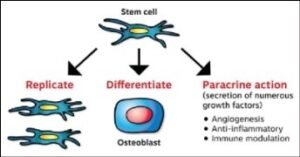
1.Therapeutic Angiogenesis – Necessary for delivery of nutrients & oxygen required for Regeneration.
2.Tissue Rejuvenation – Growth factors secreted by cells Modulate the LocoregionalMilieun. Promote self-repair of residing tissue cells. Activate the dormant local regenerative cells of damaged tissue
3.Differentiation – These cells are themselves a rich source of progentior cells Differentiate in bone or cartilage under optimum conditions

Osteogenic differentiation chondroblastic differntiation
HOW IT IS DONE
Patients are evaluated clinically, Radiologicalyby XRAY ,CT or MRI scans. Patients advised to strictly avoid smoking, drug or alcohol consumption and have a positive life style. As a day care procedure 240 ml of Bone marrow and 100 ml of fat is aspirated and processed for stem cells isolation. 2 ml of stem cells processed as per GMP is injected directly
1. Damaged Joint by intrarticular Injection under ultrasound guidance
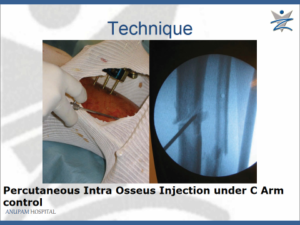
2. Intralesionalat Fracture site under fluoroscopic control
3. Intralesional in AVN under fluoroscopic control
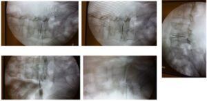
4. Intralesional administration of cells in disc/facets in LBPunder fluoroscopic control
The procedure is very simple as administration of cells can be done to target site directly Intrarticularor Intralesional under ultrasound guided or C-ARM fluoroscopic control easily

Percutaneous Intralesionaladministration at Fracture site under fluoroscopic control

Intrarticular Injection into Damaged Joint

Intralesional in AVN

C Arm images showing administration of cells in disc for disc disease LBP

C Arm images showing Intralesionali Administration of cells in facets for LBP
What to expect
OSTEOARTHRITIS OF KNEE
- WOMAC values (subsets and total) showed significant improvement from baseline over the course of six months treatment (p < 0.001).
- The pain free distance covered during a Six minute walk (6MWD) also significantly improved by 140 feet ((p<.001).
- Requirement of rescue medication decreased to once a week in 45%.
- The mean (SD) change for the percentage of painful days was 45.0 (38.7).
- 25% patients demonstrated improved cartilage thickness by at least 0.01 mm as against 15% who lost thickness at least by 0.01 mm.
- 55% patients showed improved Cell counts (below 500 microlitres) in Synovial fluid.
DELAYED AND NONUNION
- Accelerates healing 90 % patients showed quick healing when used as a supplement with first surgery where high chance of avascular necrosis and Nonunion exist e.g.-# neck femur/ talus/distal tibia
- Achieved union in 95% of cases in cases of Delayed union (when X-ray showed no callus in bridging construct or callus in stable construct in 3 -6 months)
- Healing seen within 3 months in 80 % cases of atrophic non-union, failed implants of long bones after autologous bone marrow concentrate.
- Cell therapy accelerated bone healing in the femur compared to in the tibia.
AVASCULAR NECROSIS OF HEAD FEMUR
- Significant reduction in pain in all the patients
- Back to work within 6 months after treatment.
- Avoided the progression of the disease effectively in 90 % of hips with Stage I & II and in 40 % of hips with Stage III & IV disease.
- 10 % of hips with stage 1 and II progressed to Stage III: as compared to 50 % of Control patients progressing to Stage III.
- 6 % of Stage 1 and II of the hips needed Total Hip replacement
- 60 % Stage III & IV hips needed Total Hip replacement
- Mean volume of necrotic lesion in Control group increased while volume was reduced in BM-MNC Group at 24 months
INTRACTABLE BACK ACHE
- Very useful in black disc disease
- 60% of patient’s Intractable Back Pain, Failed Spine surgeries, respond to cell application
- VAS Pain scores improved significantly and maintained for 18 months in 60 percent of subjects.
